
Microbiology Fundamentals: A Clinical Approach
3rd Edition
ISBN: 9781259709227
Author: Marjorie Kelly Cowan Professor, Heidi Smith
Publisher: McGraw-Hill Education
expand_more
expand_more
format_list_bulleted
Concept explainers
Videos
Textbook Question
Chapter 3, Problem 1VC
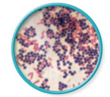
From chapter 2, figure 2.18. Explain why some cells are pink and others are purple in this image of a
Expert Solution & Answer
Want to see the full answer?
Check out a sample textbook solution
Students have asked these similar questions
Peptidoglycan on bacterial cell walls is stained by crystal violet dye and for this reason those bacteria with a
thick layer of peptidoglycan have purple appearance in gram dye/stain test.
True
False
Normal
Streptococcus pyogenes
No Spacing
Heading 1
Heading 2
Streptococcus pneumoniae
Title
you observed the growth of Streptococcus pyogenes, Streptococcus pneumoniae and
Staphylococcus epidermidis on the Horse Blood Agar (HBA) plates. The colonies and their
surroundings looked like those in the image below. Based on the content of (HBA) plates
and the specific properties of these three organisms, explain the particular appearance of
the colonies on the plates. Name and describe the three subtypes of a process that occur on
these plates.
Styles Dictate Sensitivi
Pane
Staphylococcus epidermidis
Focus
E
E
1
Figure 3. Two different types of bacterial colonies on an agar plate.
Using microbiology terms, describe fully the colonial morphology of the two colonies shown
above. A full description will include texture, transparency, color, and form (size, overall shape,
margin, and elevation).
Colony 1
Colony 2
2.
Chapter 3 Solutions
Microbiology Fundamentals: A Clinical Approach
Ch. 3.1 - List the structures all bacteria possess.Ch. 3.1 - Identify three structures some but not all...Ch. 3.1 - Describe three major shapes of bacteria.Ch. 3.1 - Provide at least four terms to describe bacterial...Ch. 3.2 - Describe the structure and function of six...Ch. 3.2 - Prob. 6AYPCh. 3.2 - Q. Device-associated infections are very common...Ch. 3.3 - Differentiate between the two main types of...Ch. 3.3 - Prob. 8AYPCh. 3.3 - Prob. 9AYP
Ch. 3.3 - Prob. 2MMCh. 3.4 - Identify seven structures that may be contained in...Ch. 3.4 - Prob. 11AYPCh. 3.4 - Prob. 1NPCh. 3.5 - Compare and contrast the major features of...Ch. 3.6 - Differentiate between Bergeys Manual of Systematic...Ch. 3.6 - Name four divisions ending in cutes and describe...Ch. 3.6 - Define a species in terms of bacteria.Ch. 3 - Archaea a. are most genetically related to...Ch. 3 - Prob. 2QCh. 3 - Suppose an argument in your city has erupted about...Ch. 3 - Prob. 4QCh. 3 - As a supervisor in the infection control unit, you...Ch. 3 - Prob. 6QCh. 3 - Prob. 7QCh. 3 - Prob. 8QCh. 3 - Bacteria and archaea have a much greater diversity...Ch. 3 - Prob. 10QCh. 3 - Bacteria have been found to change the structures...Ch. 3 - Bacterial and archaeal chromosomes are not...Ch. 3 - Prob. 13QCh. 3 - The results of your patients wound culture just...Ch. 3 - We know that bacteria/archaea and their genetics...Ch. 3 - Find the true statement about biofilms. a. They...Ch. 3 - Suggest more than one reason why bacteria may...Ch. 3 - Construct arguments agreeing with and refuting...Ch. 3 - Which of the following would be used to identify...Ch. 3 - During the cold war between the Soviet Union and...Ch. 3 - During the cold war between the Soviet Union and...Ch. 3 - From chapter 2, figure 2.18. Explain why some...
Knowledge Booster


Learn more about
Need a deep-dive on the concept behind this application? Look no further. Learn more about this topic, biology and related others by exploring similar questions and additional content below.Similar questions
- Put the following Steps of gram smear in order Create a bacteria Add crystal violet to the bacterial smear. Rinse with water after 60 seconds. Allow the smear to air dry and then heat fix it. Add gram iodine to the smear, let it sit for 60 seconds and rinse water decolorize with 95% ethanolfoe 5-10 seconds and then rise with water Blot dry with bibulous paper Add safranin to the smear and let sit for 60 seconds, then rinse with water Observe using oil immersionarrow_forwardIn Figure 5-5,a. Why do A− and B− cells, by themselves, not formcolonies on the plating medium?b. What genetic event do the purple colonies in themiddle plate represent?arrow_forwardThe below photograph shows a Gram-stained slide viewed using a light microscope set at brightfield of a clinical sample from a patient with symptoms suggesting an infection. The slide was viewed with x100 objective. What could be the most likely cause of infection? Gram positive bacteria, either rods or cocci Gram positive rod shaped bacteria or yeast Gram negative rod or cocci shaped bacteria Gram negative bacteria, either rods or cocciarrow_forward
- This is how a hanging-drop slide is prepared. Figure from Macedo, Wikimedia Commons, 2016. 2 3 4 slide cover slip vaseline concavity drop of microbiological culture CA inoculation loop 11. Which slide gives you more information, the hanging drop or the stained slide? Why do you say so? 12. Why might it be valuable to know whether a bacterium is motile?arrow_forwardExplain why some cells are pink and others are purple in Gram-stained bacterial smear.arrow_forwardExplain why some cells are pink and others are the Gram- stained bacterial smear. Add references and sourcearrow_forward
- When you interpret a Gram-stained smear, you should also describe the morphology (shape) of the cells, and their arrangement. In the figure below, there are two distinct types of bacteria, distinguishable by Gram stain reaction, and also by their shape and arrangement. Below, describe these characteristics for both bacteria: Gram positive bacterium Gram negative bacterium Morphology cocci bacillus Arrangementarrow_forwardHello, Please answer the following attached Microbiology question AND ANSWER ALL 3 PARTS COMPLETELY. Thank you. *If you actually solve all of the THREE PARTS (QUESTIONS) CORRECTLY AND COMPLETELY, I will 100% leave a thumbs up. Microbiology Question: "The attached picture is a gram stain for mycobacterium tuberculosis. What does the Gram Stain photo look like? Based on this attached picture, please explain whether it is Gram-positive or negative. Also, please explain the colors chemistry and shape. Thank youarrow_forwarda. Identify the cell shape, cell arrangement, and Gram reaction of the Unknown microbe in the image below. b. Suppose the corresponding Gram negative control were purple. Explain what step(s) of the Gram stain you would adjust and how you would adjust them to ensure the accuracy of your Unknown resultsarrow_forward
- What is the shape of the bacteria in the pictures under microscope ( Cocci or Bacilli ) and why with explanation? These bacteria types gram positive or gram negative and why ? Please compare and discuss of these bacterias the shape and gram positive or negative according to web resources. State your reasons why you think the shape and gram positivity of both bacterias Are those images OK? OR if there is something wrong in the images what is it? What is the problem? What could it be done for getting more quality images in Gram staining? What do we pay attention in Gram staining?arrow_forwardDescription Shape: Arrangement: Photo by 1000x MITPanganiban Figure 2.9. Microscopic morphology of Bacillus cereus. Description Shape: Arrangement: Photo by 1000x MITPanganiban Figure 2.10. Microscopic morphology of Staphylococcus aureus..arrow_forwardAnswer this question xi) What is the most important step in the Gram staining procedure? Why?arrow_forward
arrow_back_ios
SEE MORE QUESTIONS
arrow_forward_ios
Recommended textbooks for you

cell culture and growth media for Microbiology; Author: Scientist Cindy;https://www.youtube.com/watch?v=EjnQ3peWRek;License: Standard YouTube License, CC-BY